
Mycoplasma synoviae occurs worldwide and is one of the two most consequential avian mycoplasmas alongside Mycoplasma gallinarum, with recognized roles in variety of illnesses which includes infectious synovitis with joint and tendon-sheath exudation, upper-respiratory infections and a unique laying-hen syndrome marked by decreased production and degrading shell integrity of the eggs known as Eggshell Apex Abnormalities (EAA). Transmission of Mycoplasma synoviae occurs both vertically via eggs and horizontally through close contact, with disease expression exacerbated by co-infections (IBV, NDV and E. coli) and environmental stressors which increases respiratory and systemic involvement. MS is a major global poultry pathogen as it shows an 11% drop in daily egg production with EAA affecting up to 24.5% of eggs in controlled trial infection, underscoring direct productivity and quality losses (Kursa et al., 2019). From year 2017 to 2021 a PCR study was conducted in India which showed that Mycoplasma synoviae positivity was around 23.61% (compared to Mycoplasma gallinarum 6.43%) with 15.49% co-infection. This suggests that Mycoplasma synoviae is the most common mycoplasma burden in Indian breeder and layer systems and a persistent economic hazard (Giram et al., 2022).
MS-associated EAA has a direct influence on income and biosecurity expenses because it increases cracked and degraded eggs, increases labour costs for sorting and cleanup and decreases hatchability through higher embryonic mortality when shell integrity is compromised. EAA manifests as irregularities at the egg’s apex, including thinning, increased translucency and susceptibility to cracks. These defects lead to increased egg breakage and spoilage, directly leading to degrading egg quality and marketability.v Etiology and Transmission: Mycoplasma synoviae, belongs to the Mycoplasmataceae family and is fastidious about its culture conditions as it requires serum and NAD on modified Frey media. The pathogenicity of strains varies due to immune evasion, adhesins, sialidase activity, nitric oxide generation and antigenic diversity.
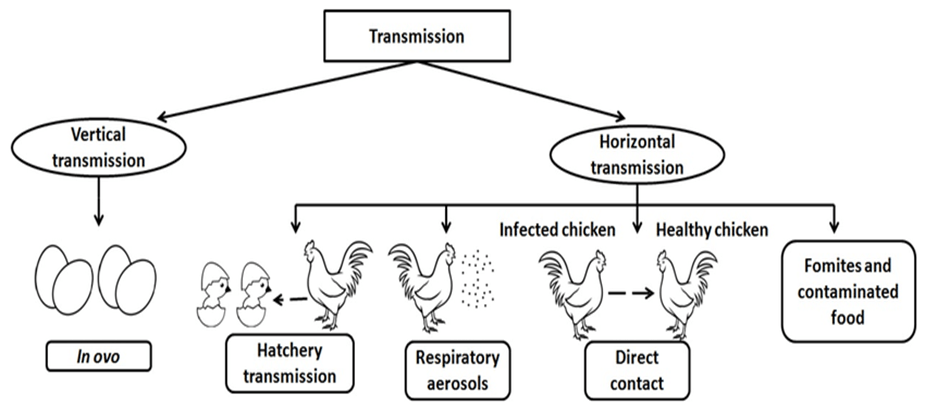
Fig. 1. Transmission of M. Synoviae
The host range of the MS infection includes chickens, turkeys, ducks, geese, guinea fowl, pheasants, quail and psittacines. Transmission occurs via both vertical and horizontal route. Vertical transmission takes place through transovarian infection, leading to early chick exposure, while horizontal transmission occurs via aerosol spread, respiratory secretions, fomites and human activity. Once introduced, the infection tends to persist, as infected flocks become lifelong carriers. Multi-age layer systems further support its persistence and contribute to episodic clinical outbreaks.
v Pathogenesis:M. synoviae primarily enters the host through the respiratory tract, with the upper respiratory mucosa serving as the initial site of colonization. With the help of specialized surface proteins and adhesions the organism attaches to the epithelial cells which help it to evade mucociliary clearance. From the respiratory tract, it can spread locally, causing tracheitis, airsacculitis and respiratory distress.In some birds, the pathogen disseminates via bacteraemia, reaching synovial membranes and joints, where it induces inflammation. This leads to synovitis, characterized by swelling, pain and lameness, often accompanied by exudation of yellowish synovial fluid. The organism may also localize in the tendon sheaths and bursae, producing tenosynovitis. Co-infections with other respiratory pathogens (e.g., E. coli, NDV and IBV) exacerbate disease severity. Chronic infections are common and affected birds may become carriers, serving as reservoirs for flock-to-flock transmission.
v Clinical Signs: Mycoplasma synoviae most commonly causes subclinical upper respiratory infections or infectious synovitis and tenosynovitis, while in layers it is also associated with eggshell apex abnormality (EAA) syndrome, characterized by thin, rough, translucent shell apices and intermittent production loss (Feberwee et al., 2009). The clinical expression of the disease is often expressed by stress and co-infections with pathogens such as infectious bronchitis virus (IBV), Newcastle disease virus (NDV) and Escherichia coli (Lockaby et al., 1998).
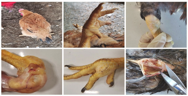
Fig.2. Dull, depressed hen, Inflammation of foot pad, hock joint and cavity filled with exudates
Affected birds may show mild respiratory involvement, including slight tracheal rales and sinusitis which are more evident under poor air quality or concurrent respiratory infections. The musculoskeletal form is marked by lameness, reluctance to walk, swelling of the hock joint, wing joints and footpads with exudative tenosynovitis of tendon sheaths and sternal bursitis. In systemic or severe cases, signs include depression, inappetence, ruffled feathers, weight loss and pale to cyanotic head parts, with occasional vasculitis and keel bursitis. Morbidity typically ranges from low to moderate, while mortality is generally low but may increase in the presence of secondary bacterial infections, wet litter, cold stress and immunosuppression.
v Post Mortem Lesions:
- Respiratory tract: Ø Mild to moderate airsacculitis with thickening, opacity and presence of turbid or caseous exudate.Ø Mucoid tracheitis and sinusitis (especially when complicated by co-infections).
- Joints and musculoskeletal system: Ø Synovitis: Swollen joints (particularly hock, wing and foot joints) with accumulation of yellow to serofibrinous exudate.Ø Tenosynovitis: Inflamed tendon sheaths filled with exudate.
Ø Sternal bursitis (breast blisters) with fibrinous to caseous material.Systemic involvement:
Ø Generalized fibrinous polyserositis in some cases, especially with secondary E. coli infection.Ø Emaciation and poor body condition due to chronic disease.Eggshell apex abnormality (in layers):
Ø No specific gross lesion in reproductive tract, but post-mortem examination may reveal rough, thin and translucent apices of eggshells in affected flocks.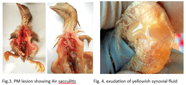
v Diagnosis: Diagnosis of MS relies on combination of clinical observation, serology, microbiology and molecular techniques. Observation of respiratory signs such as sneezing, coughing and nasal discharge, along with joint or tendon swelling indicative of synovitis or tenosynovitis and specially in layers, eggshell apex abnormalities like thin, rough or translucent apexes can be observed. However, clinical signs alone are not definitive, as they can overlap with other infections like NDV, IBV or E. coli. Serological tests, including ELISA, rapid plate agglutination (RPA) and hemagglutination inhibition (HI), are useful for flock-level monitoring, though maternal antibodies and past exposure can complicate interpretation. Microbiological isolation from choanal or tracheal swabs and synovial fluid using specialized media allows definitive identification of MS, but the process is slow and prone to contamination. Molecular methods such as PCR and real-time PCR offer rapid, sensitive and specific detection of MS DNA, even at low bacterial loads. For accurate diagnosis, a combination of clinical assessment, serology and molecular confirmation is recommended, especially in flocks showing respiratory disease, joint swelling, or eggshell defects.v Treatment Alomg with careful use of antibiotics, proper management practices and vaccination strategies are very important in Myciplasma synoviae management. Treatment typically relies on antimicrobials such as tylosin, tiamulin, doxycycline or enrofloxacin, which can reduce bacterial load and clinical signs, but complete eradication is difficult due to intracellular persistence. Widespread and indiscriminate antibiotic use has led to antimicrobial resistance (AMR) in MS strains because of these challenges, vaccination plays a central role in flock protection, lower bacterial shedding and prevent eggshell apex abnormalities in layers. v Prevention and Control:Prevention focuses on biosecurity measures, including sourcing MS-free breeders, controlling movement of personnel and equipment and minimizing stressors that predispose birds to infection. Integrated control combining vaccination, strict biosecurity, monitoring via serology or PCR and responsible antimicrobial use is essential to minimize economic losses, maintain flock health and reduce the risk of AMR development. Thus vaccination, combined with good biosecurity and management practices can control MS spread, minimizing antibiotic reliance and maintaining flock productivity. Stallen South Asia Pvt Ltd is offering a unique inactivated vaccine MS-VAC particularly against Mycoplasma synoviae. v Key Features of MS-VAC:Ø The Only Vaccine Made from highly immunogenic strains of Mycoplasma synoviaeØ High titre (1010 CFU)Ø Oil adjuvantØ High immunogenicity. Ø High safety, effective protection and field compatibility
v Duration of immunity in MS-VAC
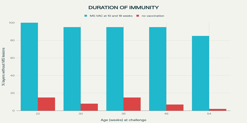
Fig. 5 Duration of immunity in MS-VAC (3 weeks after challenging with virulent MS) MS-VAC is a vaccine produced from highly immunogenic strains of Mycoplasma synoviae. The culture is inactivated and emulsified in light mineral oil, to ensure a high degree of protection after first vaccination, however the immunity is strongest and long lasting after second inoculation. Ø Clinical observation of egg laid, in vaccinated and non vaccinated commercial hens, after infection by field MS.
v Field efficacy of MS-VAC against eggshell apex abnormalities (EAA)
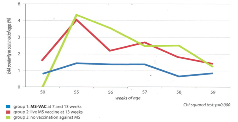
A significantly lower (p=0,000) percentage of EAA affected eggs was observed in group 1 than in groups 2 and 3 (statistically significant difference for p<0.001).Thus, MS-VAC proved to be effective in protecting commercial hens from EAA, significantly more than the competitiors, in farms infected with MS.
v Reference:
- Feberwee, A., de Wit, J. J., & Landman, W. J. M. (2009). Induction of eggshell apex abnormalities by Mycoplasma synoviae: Field and experimental studies. Avian Pathology, 38(1), 77–85.
- Giram, P., Bhutada, P., Prajapati, C., Koratkar, S. S., Patil, S., Hooda, D., Rale, V., & Tongaonkar, S. S. (2022). Percent positivity and phylogenetic analysis of Mycoplasma gallisepticum and Mycoplasma synoviae in commercial poultry from different states of India. Veterinary World, 15(7), 1843–1851.
- Kursa, O., Feberwee, A., & Landman, W. J. M. (2019). Eggshell apex abnormalities caused by two different field strains of Mycoplasma synoviae in experimentally infected chickens. BMC Veterinary Research, 15(1), 1–9.
- Lockaby, S. B., Hoerr, F. J., Lauerman, L. H., & Kleven, S. H. (1998). Pathogenicity of Mycoplasma synoviae in broiler chickens. Veterinary Pathology, 35(3), 178–190.