D. Kumar1 .M. Patidar2, A. Patidar3, S. C. Meena4, Saranaya5.R,
1, 2, 3, 4, 5Scientist RRS CAZRI Jaisalmer
Lumpy skin disease was first seen as an epidemic in Zambia in 1929. Initially, it was thought to be the result of either poisoning or a hypersensitivity to insect bites. Additional cases occurred between 1943 and 1945 in Botswana, Zimbabwe, and the Republic of South Africa. Approximately, 8 million cattle were affected in a panzootic infection in South Africa in 1949, causing enormous economic losses. LSD spread throughout Africa between the 1950s and 1980s, affecting cattle in Kenya, Sudan, Tanzania, Somalia, and Cameroon. In 1989 there was an LSD outbreak in Israel. This outbreak was the first instance of LSD north of the Sahara desert and outside of the African continent. This particular outbreak was thought to be the result of infected Stomoxys calcitrans being carried on wind from Ismailiya in Egypt. During a period of 37 days between August and September 1989, fourteen of the seventeen dairy herds in Peduyim became infected with LSD. All of the cattle as well as small flocks of sheep and goats in the village were slaughtered. Throughout the past decade, LSD occurrences have been reported in Middle Eastern, European, and west Asian regions. Lumpy skin disease is a vector-borne pox disease of domestic cattle and Asian water buffalo and is characterized by the appearance of skin nodules.The disease has been reported in several Indian states like Assam, Odisha, Maharashtra, Kerala, Karnataka, Chhattisgarh, Madhya Pradesh, and Rajasthan etc.
Susceptbile hosts
Lumpy skin disease is host-specific, causing natural infection in cattle and Asian water buffalo (Bubalus bubalis). Lumpy skin disease does not affect humans.
Causal organism
Lumpy skin disease is caused by the lumpy skin disease virus (LSDV), a member of the genus Capripoxvirus (CaPV) within the family Poxviridae. Lumpy skin disease virus shares the genus with sheep pox virus (SPPV) and goat pox virus (GTPV), which are closely related, but phylogenetically distinct. Transmission of the virus occurs through movements of cattle. Infected animals showing lesions in the skin and mucous membranes of the mouth and nasal cavities excrete infectious LSDV in saliva, as well as in nasal and ocular discharges, which may contaminate shared feeding and drinking sites.The virus persists in the semen of infected bulls so that natural mating or artificial insemination may be a source of infection for females. Infected pregnant cows are known to deliver calves with skin lesions. The virus may be transmitted to suckling calves through infected milk, or from skin lesions in the teats.Local blood-feeding insect vectors feeding on cattle can also transmit the virus. The common stable fly (Stomoxys calcitrans), the Aedes aegypti mosquito,
and some tick species of the Rhipicephalus and Amblyomma spp., have demonstrated ability to spread the LSDV.
Symptoms
- Initial symptoms – Lachrymation and nasal discharge
- Subscapular and prefemoral lymph nodes become enlarged and are easily palpable.
- • High fever (>40.50C) may persist for approximately a week.
- Sharp drop in milk yield.
- Appearance of highly characteristic, nodular skin lesions of 10-50 mm in diameter: The number of lesions varies from a few in mild cases to multiple
lesions in severely infected animals. Predilection sites are the skin of the head, neck, perineum, genitalia (Fig. 9), udder and limbs. Skin nodules may persist for several months. - Sometimes, painful ulcerative lesions develop in the cornea of one or both eyes, leading to blindness in worst cases.
- Pneumonia caused by the virus itself or secondary bacterial infections, and mastitis are common complications.
- Infected animals often recover within three weeks of treatment with anti-allergy and antibiotic medicines. The morbidity rate in LSD is 10-20%, while the mortality rate is up to 5%.
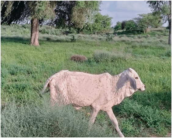
Picture: cow infected with lumpy in Jaisalmer
Measures to taken on farm in case of suspicion
- If possible, separate the suspected case(s) from the rest of the herd.
- If possible, separate the rest of the animals from neighbouring herd(s) by feeding them on the farm and avoiding communal grazing
- Disinfect your hands, footwear, and outfit using any common disinfectant and when at home/farm wash the clothes at +60 °C.
- Disinfect equipment and materials used in the affected holding.
- Contact your veterinarian for support.
Prevention
- A careful surveillance of the disease onset and spread is to be taken up at the farm level.
- Purchase of new animals that are either incubating the disease or are viraemic without exhibiting any symptoms presents a major risk of introducing the disease into a naïve herd. Introduction of new animals into herds should therefore be limited. Stock should be bought only from trusted sources. New animals should be examined and declared free of clinical signs prior to movement and on arrival, and should be kept separated/quarantined from the herd for at least 28 days
- In affected villages, cattle herds should be kept separate from other herds by avoiding communal grazing.
- Cattle should be treated regularly with insect repellents to minimize the risk of vector transmission of the disease. This measure cannot fully prevent transmission but may reduce the risk.
- Limiting vector breeding sites such as standing water sources, slurry and manure, and improving drainage in holdings are sustainable, affordable and environmentally friendly ways of reducing the number of vectors on and around cattle.