Calving is the most critical step for completion of gestation. There are many unrelated complications that can occur during gestation in dairy animals. In this article we will highlight the most important and highly prevalent gestational disorder which effects the farmer’s output in terms of foetal survival.
- Uterine Torsion: Rotation of uterus along its longitudinal axis is defined uterine torsion. It usually occurs in pregnant animals. Among buffaloes, it accounts for 50-70 % single largest cause of maternal dystocia. Possible etiological factors are poor growth of / weaker broad ligaments, lesser placental fluid, decrease uterine tonicity, uncoordinated or boisterous fetal movements and /or strenuous exercise of pregnant animal. Due to presence of large rumen in the left side of abdomen usually right side twisting is more common. Major clinical symptoms are:
- Pain: usually torsion occurs in the last trimester of pregnancy i.e from 6-7 months onwards. Animal will feel pain and it expresses it by cyclic activeness of sit and stand again and again. Animal will lie down on the ground and kick her abdomen. Due to the uterine twisting animal will show pain in caudal abdominal area.
- Straining: If torsion occurs towards the last month of gestation then we can also see frequent straining in the animal without futile results.
- Anorexia: Feed intake of the animal will decrease. Due to this pain in the caudal abdominal area animal will take less feed and water which results in dehydration and scanty faecal output.
Diagnostic evaluation of the condition should be done in the initial stages. Delayed case of uterine torsion will have grave prognosis. During torsion of uterus twisting of blood vessels also occur, which result in haemorrhages in the uterus. This blood due to gravitational action will flow downwards and starts to clot and leads to adhesion formation from the ventral abdominal portion. These adhesions will start aligning upward from the ventral abdomen. Early the case is diagnosed lesser will be the uterine adhesion and more are the chances for detorsion to be achieved easily. If the case is delayed there is more adhesion so more chances of uterus to be torned out during detorsion, hence the prognosis is grave. The success of management of the condition lies in the correct and timely diagnosis and early referral to referral centres. In cases presented within 24 h of the problem, rolling is often successful in correction of the problem. So it is concluded that early presentation to referral centres culminate in successful management of the condition with high dam and fetal survival.
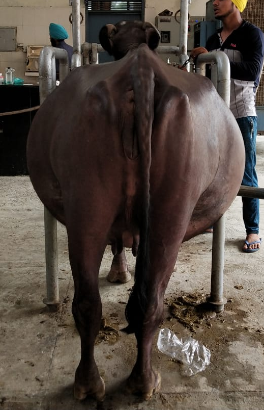
- Hydrallantois: Hydrallantois (hydrops of the allantois) is a clinical condition that occurs during the last trimester of pregnancy and is characterized by larger than normal accumulation of allantoic fluid. There is bilateral abdominal enlargement with uterus filled with about 40 to 160 litres of fluid. Abdominal enlargement looks like barrel shaped from behind, completely stretched belly with fluid thrills can be palpated while tapping and teats are like tucked up to the belly.
Due to abnormally large amount of allantoic fluid accumulation in the uterus, it will push rest visceral organ towards anterior side and hence the correlated symptoms will follow. Major clinical symptoms:
- Anorexia: Symptoms like less feed intake due to uterus pushing rumen and lesser space is available for feedstuff. So the animal will be anorectic and due to lesser feed intake impaction will occur. Hence the animal will pass scanty black faeces and due to rectal straining rectal mucosa will also get abraded. So the faeces will have mucoid rectal debris also. Hence no rumination will occur.
- Dyspnoea: This filled uterus will also put pressure on diaphragm which hinders the complete expansion of pleural parenchyma and thus the animal will have difficulty in breathing also accompanied with open mouth breathing. The extent of dyspnoea will increase with the advancing gestation, if timely correction of problem is not done.
- Bizarre conformation: Due to abnormally large amount of allantoic fluid accumulation in uterus animal will be having a bizarre conformation with abdomen completely stretched up, fluid thrills on tapping the abdomen and teats tucked up on the belly region. When looked from behind animal’s abdomen will appear as barrel shaped i.e. bilaterally distended abdomen.
- Fluid accumulation: This huge amount of fluid accumulation will occur in a period of 10-15 days. Initially the appetite of animal will be normal and suddenly it reduces as the fluid accumulation fastens. Normally the allanoic fluid present in the uterus is around 7-20 L and in this case it increases maximum to 200 times i. can accumulate up to 200 L.
Hydroallantois is more common in bovines and rarely occurs in mares, ewes and goats. It accounts for about 85-90% of the dropsical conditions in bovines. The exact cause of this disease is uncertain and but scientist has speculated that it occurs due to diseased uterus, non-functional caruncles and enlarged placentomes. Adventitious placentae are commonly present and there may also be a deficient number of caruncles. A reduction in the number of cotyledons has also been associated with it. Others factors that contribute to the disease condition are increased permeability of the chorio-allantoic membrane, decreased sodium transport across the chorioallantoic membrane, fetal renal/hepatic diseases, hormonal imbalance, multiple foetuses, uterine torsion and/or twisting of the umbilical cord, deficiency of vitamin A thus causing decreased endometrial resistance which further contributes to hydrallantois.
The slow removal of excessive allantoic fluid should be practised to prevent the animal from hypovolemic shock. The antibiotic, antihistamine and intravenous fluid should be done for faster recovery.
- Mummification: Fetal mummification is characterized by complete resorption of fetal fluid during gestation without any bacterial contamination which leads to wrapping of uterus around the fetus.There are certain number of events that must be present in order for fetal mummification to occur: 1) the fetus must die after the complete ossification of bones otherwise autolysis occurs rapidly and soft tissues are digested and absorbed through the endometrium, 2) rapid resorption of uterine and fetal fluids, 3) no oxygen in the uterus until the mummification process is complete, and 4) no bacterial invasion inside uterus, here closed cervix prevent the entry of putrefactive organisms present in the vagina and vestibule. Major clinical symptoms are:
- Over-gestation: Due to dead fetus and no signal of initiation of calving the dam will retain the fetus till she is examined by the veterinarian. Dam may take more time than normal gestation period. Gestation period of the dam may be extended up-to one month.
- No change in feeding behavior is noticed. As there is no infection inside uterus and no septicemia so there will not be any decrease in feed intake or other GI problems.
- Mummification can occur at any time of gestation so we can notice that from one point of time in gestation there is no size increase in the abdominal area due to fetal growth.
- No putrid discharge or normal cervical seal will come out from vagina.
The entire process of mummification will occur in several weeks, depending on the age of the fetus at the time of death. After complete resorption of fetal fluid, fetal membranes and uterine wall adhere closely to the fetus, and whole mass becomes blackish and leathery in appearance. It may or may not have some exudate resulting from the degeneration of red blood cells.
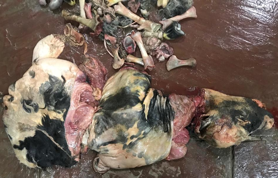
- Maceration: Fetal maceration is the fragmentation of a fetus that has died after ossification of the fetal bones, which led to putrefaction and digestion of soft fetal tissue due to bacterial invasion. Non-delivery of a dead fetus could be due to partially dilated cervix and abnormal presentation of a partially dry fetus. Fetal maceration may occur at any stage of gestation. Sometimes the disintegrated fetus parts and bones may be retained in the uterus for prolonged periods necessitating surgical removal. Major clinical signs are:
- Generalized dullness: Bacterial contamination will led to generalized weakness and dullness in demeanor. Sunken eyes with loss of contour are common finding.
- Anorexia and pyrexia: Due to bacterial invasion and septicemia, animal will be anorectic. Septicemia will also lead to pyrexia due to generalized body infection.
- Vaginal discharge: Due to bacterial colonization inside the uterus and following putrefaction, there is foul smelling sometimes blood tinged vaginal discharge comes out from uterus.
- Frequent straining: Open cervix and putrid vaginal discharge indicates initiation of calving but due to incomplete cervical dilation progression is hindered with no result. Animal will strain frequently with no outcome.
It is necessary to get the animal checked by qualified veterinarian to diagnose all the above mentioned conditions and to get a remedy at the earliest to save the life of animal.
Ankit Kumar Ahuja, M Honparkhe, S S Dhindsa, Prahlad Singh
Department of Veterinary Gynaecology and Obstetrics, Guru Angad Dev Veterinary and Animal Sciences University, Ludhiana, Punjab.