Arsha Shaji, Dipti Nain, Surajit Das
PhD scholar, Animal reproduction, Gynaecology and Obstetrics,
ICAR-National Dairy Research Institute, Karnal-132001
Abstract
A Doppler ultrasound is an imaging test that uses sound waves to show blood moving through blood vessels. Doppler ultrasonography serve to be a promising tool for solving the hurdle of earliest diagnosis of pregnancy in several domestic animals.For the purpose of deciding whether to rebreed or cull non-breeding females, identification is crucial.Pregnancy can be detected early enough to prolong a female’s productive period.Colour Dopplercan also be used for studying follicular dynamics, uterine changes, and pathological changes in the reproductive tract in all the domestic animals. It is widely used to detect the blood supply in various genital organs to easily predict the pregnancy.
Introduction
The objective of pregnancy diagnosis is to determine the pregnancy status with100% accuracy, no false positives, no false negatives and to determine the pregnancy as early as possible. It is done tobe able to determine the age ofthe conceptus, determine the viability and sex of the fetus and get the results early as possible. Pregnancy diagnosis is a critical step in the reproductive management of any animal production system. Identification of non-pregnant females is important for decision-making regarding rebreeding or culling of those animals. The earlier the pregnancy can be diagnosed, the earlier decisions can be made; thus, an early, accurate, and feasible pregnancy diagnosis method is essential for optimizing reproductive performance. The target calving interval for cattle and buffaloes should be 365 and 405 days, respectively but the existing ones are 460 days and 530 days, respectively (Annual report NDRI 2014-15). In crossbred cows, if the calving interval is reduced by 1 month due to early diagnosis of pregnancy, then we can expect a gain of around 132 productive days.To achieve a desirable calving interval, the foremost requirement is the identification of non-pregnant cowspost-service so that they can be re-bred at an early date. Although there are several techniques such as rectal palpation, progesterone assay, and early conception factor for early pregnancy diagnosis, the efficiency of pregnancy detection with these available techniques varies from 30-93%.However, the colour doppler ultrasonography serve to be a promising tool for solving the hurdle of earliest diagnosis of pregnancy in several domestic animals.
Methods for pregnancy diagnosis
| Methods | Drawbacks | Efficiency | Reference |
| Non-return to oestrus | Time-consuming,labor-intensive | 30%- 50% | Leven et al.,2013 |
| Progesterone assay | Costly, skill, low specificity | 24 day-93.1% | Pieterse et al.,1990 |
| Pregnancy-associated glycoproteins | Costly, need laboratory facilities | 28 day-93.9% | Romano et al.,2010 |
| Early conception factor | Lack specificity | 9 & 15 days- 55% | Whisnant et al.,2001 |
Colour Doppler Ultrasonography
An ultrasound beam meets a moving object the reflected ultrasound is either of increased or decreased frequency, depending upon whether the motion is towards or away from the transducer.
Blood towards probe: +ve doppler shift
Blood away from the probe: -ve doppler shift
Doppler system ultrasound production
Colour-mode scan lines are emitted from the transducer. Echoes from stationary signals are removed by an echo canceler. The autocorrelator computes the average velocity from each group or packet of echoes by a process called phase-shift autocorrelation. The resulting data are used to indicate the direction and intensity of blood flow by variation in the color pixels along each scan line.Colour Doppler ultrasonography is a promising tool for diagnosis of early pregnancy. It can also be used for studying follicular dynamics, uterine changes, and pathological changes in the reproductive tract. Colour Doppler can be used in male reproductive system also inorder to ascertain its proper functioning, abnormalities etc.
Changes in blood flow during the estrous cycle in cow
Blood flow surrounding a CL increases in response to PGF2α released from the uterus 16-17 days after oestrus, initiation of luteal regression occurs. A decrease in blood flow in the CL is observed during luteal regression. Detectable blood flow is maintained around a dominant follicle during follicular deviation. However, unselected follicleswill have diminished blood flowand become atretic follicles. After the LH surge, blood flow within the follicular wall of a preovulatory follicle rapidly increases. During CL development, a gradual increase in blood flow surrounding theearly CL will happen simultaneously with an increase in plasma progesterone level.
Pregnancy diagnosis in cows using Colour Doppler ultrasonography
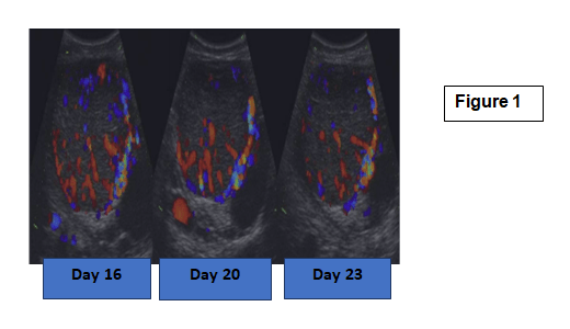
In cows, blood flow surrounding the CL decreases during CL regression, and monitoring of blood flow can be used to diagnose pregnancy. Images of CL with active blood flow at days 16, 20, and 23 of the estrous cycle in a pregnant cowshown in (Fig.1). Here, it is evident that blood flow does not change substantially over these days.
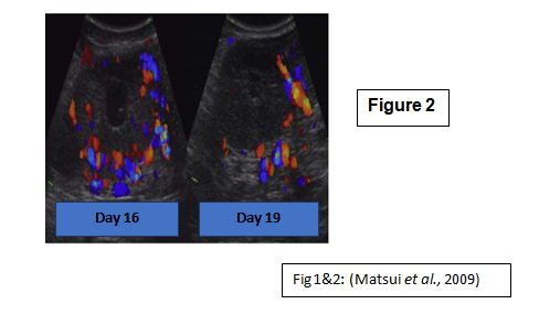
Blood flow clearly decreased at 19 days after ovulationin non-pregnant cattle and then oestrus occurs followingregression of the CL observed within 3 days after adecrease in blood flow surrounding the CL (Fig.2). These findings indicate that the CL had intiatedregression when adecrease in blood flow was observed. Therefore, a decreased blood flow surrounding the CL at 19–21 days after ovulation and AI may be an indicator of a non-pregnant cow.
Color Doppler ultrasonography for early diagnosis of pregnancy in buffalo
It is conducted in buffalo because of the limited capacity to detect estrus because of the lack of behavioral symptoms of estrus in buffalo, seasonal alteration in exhibition of estrus symptoms, and prolonged calving intervals.Determination of pregnancyin buffaloes is done with high accuracy (80.4%) at Day 17, and maximum accuracy (96.4%) at Day 21 post-timed artificial insemination using color doppler ultrasonography. The concentration of progesterone reported to be positively associated with the scores of luteal blood flowparticularly at Day 17 and 21 post-TAI. This ensures early re-synchronization of estrus in non-pregnant buffalo, with a fortnight earlier diagnosis compared with early pregnancy diagnoses conducted using trans-rectalultrasonic procedures.
Early pregnancy diagnosis in ewes using color doppler ultrasonography
In ovine, even though pregnancy might be recognized as early as 17-19 days post-breeding using B-Mode ultrasonography, but capable to predict it at least 25 days after oestrus for accurate and reliable results. Females bearing a CL with score 2 or greater were apparently considered to be pregnant. Using color doppler ultrasonography to distinguish between pregnant and non-pregnant animals is considered to be a proven tool that was highly efficient starting from 17 days after breeding in ewes.
Pregnancy diagnosis in Goats
In goats, the luteolysis phase begins from about Day 17 of the estrous cycle. During the luteolysis phase, the luteal blood flow is more sensible, which enables the detection of minor alterations in the progesterone values when compared with the luteal tissue area. Animals
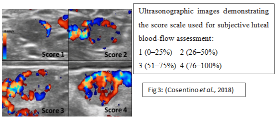
| Fig 3: (Cosentino et al., 2018) |
bearing at least one CL, with or without a cavity, with a score ≥ 2 were considered pregnant (Fig.3). Color Doppler can be judged as a reliable tool for pregnancy diagnosis in goats as early as Day 21 post-breeding, while only from Day 23 by using B-mode ultrasonography.
Pregnancy diagnosis in Mare
Color Doppler techniques could be effective in detecting a regressing CL before any decrease in circulating progesterone, or changes of a B-mode image or uterine/cervical tone become apparent in the mare. Color Doppler ultrasonography might be particularly useful for identifying individual mares where luteolysis has been initiated, given that CL blood flow decreases more rapidly after the initiation of luteolysis than CL area which is assessed by B-mode ultrasonography (17.3% per day compared with 7.7% per day)(Borganet al., 2016).
Pregnancy diagnosis in mare by assessing uterine blood flow
The blood flow pattern to the equine uterus is dependent upon the stage of the reproductive cycle, and the degree of blood supply. The vascularization of the right and left uterine horns of cyclical mares during initial pregnancies, observed that on days 11 and 15 to 29 of gestation. The uterine blood supply increases from second week of pregnancy in mares following conceptus-induced endometrial vasculogenesis. There is a marked increase in uterine blood flow between days 11 and 17, which coincides with rapid conceptus expansion. From day 11 a gradual increase in the volume of blood flow in both uterine horns during the embryonic mobility phase and a higher speed in blood flow in the uterine horn of fixation of the conceptus, in relation to the opposite side (Fereria et al., 2015).
Conclusion
Doppler ultrasonography is a technology that has potential for overcoming the challenge of early pregnancy diagnosis in a variety of domestic animals.Color doppler approach is made more crucial by the fact that the assessment of uterine content echogenicity, in association with a functional CL was not sufficiently accurate to reach an earlier pregnancy diagnosis.It was made possible by assessment of corpus luteum blood supply by using color doppler ultrasonography.