Prof. Dr. R.N. Sreenivas Gowda
Former and Founding VC, KVAFSU, Bidar, Former Director, IAH&VB, Bangalore, Former Prof. and University Head, Dpt. of Pathology, Veterinary College, UAS, Bangalore
Introduction
In recent decades, intensive poultry production has faced both positive and negative consequences. Although there are several nutritional factors causing production problems, the bacterial and viral infections largely affect the reproductive organs or create severe health problems and production loss and their quality. The pathogens with a tropism for the reproductive organs or those that cause debilitating health conditions demonstrate mild to severe adverse effects on the egg production process. Multiple factors, mainly including host species, strain of the organisms, any disease of poultry can adversely affect egg production and quality either directly by having effects on the reproductive system, or indirectly, by affecting the health of the bird. The entire farm economics directly depending on the egg production, any deviation in health jeopardise the farmer’s economy.
Several factors can affect egg production of non-infectious origin. Among these factors are age, nutrition and water supply, environmental conditions such as light and stress, and processes such as molting, poor ventilation (increased noxious gases) in poultry establishments and nutritional errors comprising of inadequate levels of energy, protein or calcium and Vit.D, Presence of mycotoxins such as aflatoxin, trichothecene, and ochratoxin can cause a drop in egg production. This is called as nutrition-based health (NBH). Therefore, it is so important to supply laying hens nutritionally balanced toxin free layer /breeder feed as per the breed, age and season for optimal egg production.
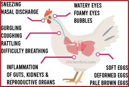
Among infectious diseases, respiratory infections cause air sacculitis and in turn, infect the ovary and oviduct (Lung- oviduct axis). Diseases such as avian influenza, Newcastle disease, Infectious bronchitis, and Infectious Laryngotracheitis (ILT)and Coryza severely affect the egg production and quality of eggs. Egg drop syndrome ’76 can cause flocks to lay shell–less eggs
The main production losses can occur from various infectious agents such as bacterial and viral agents. This paper deals with such infectious agents that causes egg production loss and the mechanism of damage to oviduct in causation of production loss.
- Bacterial Infections that Induce Ovary and /or Oviduct Disease
There are many bacteria that cause infectious diseases in laying hens have systemic effects on egg production (Table1).
Table1. Adverse effects on egg production and quality observed in different Bacterial diseases.
| Bacterial Infections | Egg production loss(%) | Pathologic lesion |
| Colibacillosis ( coli) | <10% | Oophoritis and salpingitis |
| Salmonellosis (Salmonella enterica enterica serovar Enteritidis) | %-10% | Regression of ovarian follicles |
| Fowl cholera (Pasteurella multocida) | <10% | Hyperaemic ovaries |
| Infectious coryza (Avibacterium paragallinarum) | 10-40% | Ovarian regression |
| Mycoplasma gallisepticum (Mg)and Synoviae(Ms) | >20% | |
| Ornithobacterium rhinotracheale(ORT) Infection | 10-15% |
- Colibacillosis
Escherichia coli is a common bacteria found everywhere and appear as low pathogenic or highly pathogenic in nature. This bacterium cause both primary and secondary infection characterized by coli septicemia, hemorrhagic septicemia, coli granuloma, air sac disease, swollen head syndrome, venereal Colibacillosis, cellulitis, peritonitis, salpingitis, osteomyelitis, yolk sac infection and enteritis. In all viral respiratory disease, it causes secondary infection causing air sacculitis.
Avian Pathogenic Escherichia coli (APEC) is often isolated from avian species This belongs to the Enterobacteriaceae family. E. coli is present in the gastrointestinal tract of most animals and it is excreted in high amounts through feces. After intake, its colonization in the trachea, caecum and oviduct takes around 21 weeks.
Transmission can occur through contact with infected birds or through the intake of water and feed contaminated with feces, as well as via the inhalation of agents from dust and bedding materials. Transmission can also occur when oophoritis and salpingitis develop in laying breeders, prior to the formation of the eggshell, or after it has been formed while passing through the cloaca.
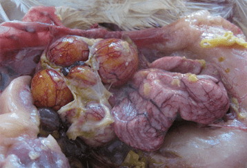
Fig.1.Egg peritonitis yolk material attached to ovarian follicles through strands.
Inflammation in the oviduct due to APEC results in the reduction of egg production and sporadic mortality. Exudate, which accumulates with the inflammation that occurs as a result of egg peritonitis causes formation of egg yolk that coagulates in the body(fig1). In addition, coli -septicemia, which affects egg production, can often be seen in young laying hens, but rarely in mature fowls.
- Salmonella Infection
These are infections caused by Salmonella Gallinarum (S. Gallinarum) and Salmonella Pullorum (Pullorum), and include Pullorum disease (PD), Fowl typhoid (FT) and infections of chicks and hens that are characterized with septicemia. Adult fowls are prone to fowl typhoid, while young fowl are prone to Pullorum disease.
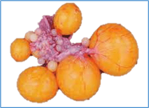
The transmission sources of Salmonella Gallinarum (S. Gallinarum) are from hatcheries, feed and rats in poultry houses. On the other hand, Salmonella Pullorum (S. Pullorum) transmission can occur within 48 hours of hatching, in which case shell penetration and feed contamination occur at a lower rate. S. Pullorum localizes in the reproductive tract of layers, and more densely in the ovary and oviduct with sexual maturation and cause severe damage to ovarian follicles (fig,2).
The most common lesions are amorphous and cystic follicles as small nodules or regression of ovarian follicles and can be seen in chronic infection. In this case, the oviduct fills with a caseous exudate, causing the dysfunction of the ovary and oviduct, thus leading to peritonitis and loss of egg production.
Fig.2 Normal and regressed ovaries with atrophic follicles (Source: Shivaprasad 2000)
- Fowl Cholera (FC)
This is a septicemic disease of poultry with high mortality and morbidity rates, caused by Pasteurella multocida (P. multocida) of the Pasteurellaceae family. Adult chickens are more prone than young fowls and broilers are more resistant to the disease than layers, resulting in deaths at higher rates in laying hens.
Transmission occurs through the digestive tract, respiratory tract, skin and conjunctiva, and is particularly transmitted through the feces or oral/nasal discharge of animals that have recovered from the infection.
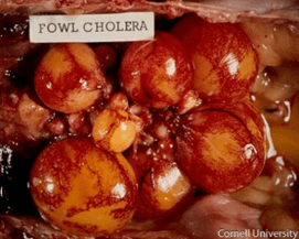
The ovaries are infected cases of acute cholera in laying hens. Matured follicles take on a flabby and densely vascularized appearance, and the follicular content is released into the peritoneum as soon as the follicles rupture. The stroma of unmatured follicles and ovaries are hyperemic, which leads to a decrease in production in laying hens.
Fig.3.Fowl cholera Hyperemic ovaries (Source: Cornel University)
- Infectious Coryza
Chickens are natural hosts of the agent Avibacterium paragallinarum (A.paragallinarum) . The disease is characterized by a swelling around the eyes and face. The agent is transmitted through secretions and excretions. Transmission can also occur through the exchange of machinery/equipment between farms, and also by personnel. Morbidity of the disease is 80–100%, while mortality is around 10%. It causes a 10–40% decrease in egg production. Feed and water consumption is usually decreased resulting in a drop in egg production.
- Mycoplasma Infection
Mycoplasma synoviae (MS) and Mycoplasma gallisepticum (MG) are the cause of mycoplasma infections, for which chickens are natural hosts. MG causes chronic respiratory infections (CRD). The primary symptoms are coughing, panting, slight opening of the beak and reduction in feed intake. Decrease in egg production, co-inflammation of the cornea and conjunctiva, facial edema and tear secretion are clinically apparent. The infection spread through “lung-gut -oviduct axis”. Oviduct thickening and salpingitis in laying hens are considered to be causes of decreases in production.
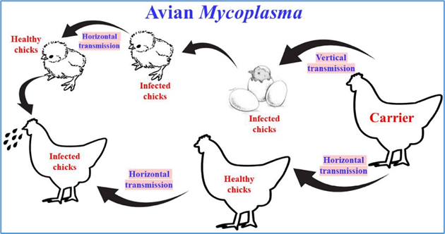
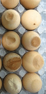
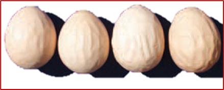
Chicks that hatch from the eggs of infected birds play a significant role in lateral transmission. The most significant route is transmission through eggs. Vertical transmission through infected eggs is observed. MS infection is seen in chickens older than 4 weeks of age. It is usually an upper respiratory tract infection. Strains isolated in recent years were frequently isolated from flocks with decreased egg production and egg defects. The agent causes the eggshell to become thinner, to lose opacity and to develop a rough surface. Thus, eggs tend to crack or break more easily. The agent causes decrease in egg production and more than 10% of eggs to be unfertilized. Mainly the apex of the shell gets damaged(fig.3).
Fig 4.Vertical and horizontal spread of Mycoplasma infection, apex damage in eggs (Source: Poultry Science Volume 102, Issue 5, May 2023, 102553)
The joint strains cause inflammation in the joints and tendons. The oviduct strain causes eggshell abnormalities (EPS), which can lead to increased breakage and an indirect and direct reduction in egg production(Fig.4).
- Ornithobacterium rhinotracheale(ORT) Infection
ORT is a contagious, fatal respiratory disease that causes growth deficiency. Its natural hosts are chickens and turkeys. It can be transmitted vertically, but also horizontally through aerosols or drinking water. The agent can be isolated from the ovary, oviduct, hatching eggs and unfertilized eggs. It affects production in commercial layers, and produce increase number of eggs of smaller size than normal and changes in shell quality are among the clinical symptoms of the disease.
- Viral Infections of Ovary and/or Oviduct Disease
Table 2. Adverse effects on egg production and quality observed in different viral diseases.
| Viral disease that affect egg production | Drop in egg production(%) | Effect on Egg quality | Lesions in reproductive organs |
| Egg drop syndrome76 | 10-40% | Soft shelled or shell less | Uterine edema, Cystic lesions in oviduct, inactive ovaries |
| Infectious Bronchitis (IB) | Up to 70% | Misshapen eggs, rough and thin shells, eggshell discoloration, and watery albumen, eggshell apex abnormalities. | Ovarian regression, atrophied oviduct, Cystic lesions in the oviduct |
| Infectious Laryngo- treacheaitis (ILT) | Up to 58% | Blood stained eggs | |
| Newcastle disease | Up to 100% | Decreased shell thickness, soft shells, spotted shells, and decreased albumen height | Small and flaccid oviducts and inactive ovaries |
| Avian Metapneumovirus (AMPV) Infection | Up to 60% | Soft or thin-shelled eggs | Ovarian hemorrhages, inspissated yolk in shell gland and regression of ovary and oviduct |
| Avian Influenza (AI) | Up to 100% | Misshapen, discolored, and fragile eggs | Salpingitis |
| Avian Encephalomyelitis (AE) | Up to 75% | – | |
| Avian Hepatitis E Virus (HEV)infection | Up to 45% | – | Oviduct regression |
| Marek’s disease | Up to 5% | – | Ovarian tumors |
| Leucosis | Up to 2% | – | Ovarian tumors |
1.Egg drop syndrome- 76 (EDS-76)
Egg drop Syndrome virus(EDSV) belongs to the genus adenovirus of the Adenoviridae family. Clinically the birds appear normal but produce egg discoloration and production of thin-shelled, soft-shelled, and shell-less eggs characterized the early outbreaks reported in the 1976. Hence the name EDS-76. Production of soft-shelled and shell-less eggs and leads to a sudden drop (10-40%) in recorded egg production or a failure to achieve a normal peak in production.
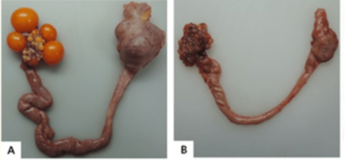
Ducks and geese are believed to be the natural hosts, but disease outbreaks commonly impact laying chickens. A drop in production and production of abnormal eggs are commonly seen in naturally occurring outbreaks(fig.5).
Naturally occurring outbreaks usually lasted 4 to 10 weeks, with a fall in egg production of up to 50%. Unlike IB no adverse effects on the internal egg quality of the eggs. Research investigating the pathogenesis of EDSV infection established the pouch shell gland as the primary site of viral replication, which was associated with the production of abnormal eggs. The lesions were minimal in mature chickens; however, uterine edema was observed. It is believed that pathological changes in the uterus of the infected bird may have interfered with the proper formation of the eggshells.
Fig 5. Gross Lesions EDS ’76. A; Normal hens’ reproductive system B; Inactive ovaries and shrunken oviduct and abnormal thin shelled eggs. (Source: From Wikipedia)
2. Infectious Bronchitis (IB)
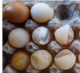
The causative agent of IB, is a gamma coronavirus (IBV) in the family Corona viridae, which exists as multiple heterologous strains. Mainly IBV initially infects the respiratory system of chickens but some strains have genital tropism. The extent of the reproductive disease may differ with the bird’s age and the strain of the causative virus. In layers, infection at peak production leads to a severe fall in egg production, accompanied by a reduction in shell quality and a watery albumen (fig,6). Alsoimpacted oviducts, ruptured ova, internal layers, and cystic right oviducts are often a result of early IB virus infection. Infectious bronchitis is also known to affect shell pigmentation. Uniformity of pigmentation in brown eggs is poor. Pale eggs can appear 2 to 5 days after exposure to the virus. The production of pale or inconsistently colored eggs could be contributed by IBV-induced lesions in the uterus, which decrease the deposition of the main eggshell pigment, protoporphyrin IX. The glandular hypoplasia of the magnum, induced by IBV infection, could result in a reduction in the synthesis of albumen proteins and, consequently, a watery albumen and reduced egg weight.
Fig 6. IB: Clinical signs, wrinkled eggs and watery yolk
Infection of female chickens within the first few days of life can cause detrimental lesions in the developing reproductive tract. Cystic dilatation in the oviduct, which has been detected in false layers with a reduced peak in egg production, could be a consequence of IBV exposure at one-day old.
3.Avian Metapneumovirus (AMPV) Infection
Avian Metapneumovirus belonging to the genus Metapneumovirus of the family Pneumoviridae . The term turkey rhinotracheitis (TRT) has been used to describe clinical respiratory disease in turkeys. In particular, manifestations suggest upper respiratory tract(URT)infection, include snacking, rales, sneezing, nasal discharge, foamy conjunctivitis, and swollen infraorbital sinus. Milder clinical signs are usually appreciated in chickens; however, swollen head syndrome (SHS) is thought to develop as a result of secondary bacterial infections.
Infection in laying birds usually contributes to a decrease in egg production and to reduced egg quality, soft shelled or thin shelled eggs. Lesions identified in the oviduct and ovary, including inspissated yolk or albumen in the oviduct, ovarian hemorrhages, and regression of ovary and oviduct. The pathogenicity of AMPV for the chicken’s oviduct was shown to be higher in the uterus than in the magnum and isthmus, which suggests a different susceptibility of epithelial cells for the three parts of the chicken’s oviduct.
4. Newcastle Disease (ND) / Ranikhet Disease(RD)
Newcastle disease virus (NDV), a RNA virus belonging to the Paramyxoviridae family. In nature, NDV primarily infects a wide variety of avian species and represents and ND is one of the most important poultry pathogens. Newcastle virus is classed into pathotypes velogenic, mesogenic and lentogenic. The viscerotropic velogenic strains cause high mortality and enteritic lesions. Neurotropic velogenic strains also cause high mortality with respiratory and nervous signs (fig.7).
Respiratory and nervous manifestations with an elevated mortality are commonly observed after the infection of non-vaccinated birds with virulent NDVs. A decreased egg production was reported in vaccinated layers challenged with a virulent ND.
Fig.7. Newcastle disease:Typical nervous tortacolis twisting of the neckand soft shelled rubbery eggs
The production of abnormal eggs, including soft-shell eggs and spotted-shell eggs are common. In the challenged specific-pathogen-free (SPF) hens demonstrated small and flaccid oviducts and inactive ovaries during necropsy examination. The uterus was reported to be the main target part of the oviduct for both live vaccine and virulent strains, which may explain the changes observed in the shell quality. The drop-in egg production was thought to be related to a decreased serum phosphorus level as a consequence of ND-induced kidney damage. Furthermore, the lower calbindin-D28k (CaBP-D28k), a calcium-binding protein, mRNA expression in the uteri of NDV-infected hens was suggested to have a role in the change in shell quality. The HI anti-NDV antibody titer levels showed a high correlation with protection against the negative effects on egg production.
5. Avian Influenza (AI)
Avian influenza viruses (AIVs) are classified as members of the genus Influenza virus- A of the family Orthomyxoviridae. Serological reactions to the surface glycoproteins, haemagglutinin (HA) and neuraminidase (NA), have been used for the subtyping of AIVs and, accordingly, sixteen subtypes of HA (H1–16) and nine subtypes of NA (N1–9) have been recognized. Influenza viruses are notorious for antigenic variation and hundreds of different subtypes exist because of antigenic drift and shift.
A large variety of avian species, both domestic and wild birds, have been shown to be susceptible to natural infections with AIVs. Poultry production has incurred huge impacts as a result of frequent outbreaks of either highly pathogenic avian influenza (HPAI) or low pathogenic avian influenza (LPAI).
The clinical signs that accompany infections with AIVs vary widely depending on the host species, age, immune status of the host, and the virus subtype involved. In chickens, for example, LPAI viruses developed disorders with mild to severe manifestations in respiratory, digestive, urinary, and reproductive systems. A sudden increase in mortality, up to 90%, may be the only indication of infection with HPAI viruses. Losses from reduced egg production have been encountered as a result of AIV infections.
Egg yolk peritonitis and salpingitis with edema in the oviduct are commonly observable lesions are the cause for egg production loss and shell quality. The significance of the widely distributed H9N2 as a primary pathogen in laying birds, infection cause a long-term decline in egg production. The virus replication-induced lesions in the infundibulum limits the reproductive functionality of the bird is the cause for drop in egg production.
6.Infectious Laryngotracheitis (ILT)
The causative agent for ILT is Gallid herpesvirus type 1 (GaHV-1), belongs to the genus Iltovirus, subfamily Alphaherpesvirinae of the Herpesviridae family . ILT is an upper respiratory disease of chickens, which is characterized by mild or severe respiratory manifestations accompanied by production losses. Like other members of the Herpesviridae family, GaHV-1 can establish latency in clinically recovered birds. Stressors such as shifting, rehousing, and an onset of laying could trigger the re-excretion of the virus in recovered chickens. A rapidly spreading ILT outbreak in a multiage (from 40 to 107 weeks) laying farms resulted in a decrease in egg production (up to 58%) and a slight increase in mortality. The gross and microscopic lesions were confined to the respiratory tissues; it was reasonable to infer that the fall in egg production was a secondary effect to the impaired heath status of the infected bird.
6.Marek’s Disease
Marek’s disease virus (MDV) cause a lymphoproliferative disease in chickens. MDV caused by cell-associated alphaherpesvirus, which was recently assigned to the genus Mardivirus within the family Herpesviridae. Live-vaccine-based control of MD is heavily practiced in laying flocks. Although clinical disease is not always apparent in infected flocks, a subclinical decrease in growth rate and egg production may be economically important. Once MDV is established, the majority of laying hens are likely to become infected regardless of any management. Longer cohort duration of virus damage the ovaries in laying hens result in greater egg production loss. The economic burden of MD comes from both direct losses from hen mortality and morbidity and indirect losses (e.g., egg production loss), caused by industry wide use of vaccines and control measures.
7.Avian Leukosis
Avian leukosis virus (ALV) infection of chickens is a substantial cause of economic losses because of tumor-associated mortalities and negative effects on production. ALV is the type species of the genus Alpharetrovirus within Retroviridae family. ALV shedding into the eggs was associated with lower egg production, delayed onset of laying, small-sized eggs, thin-shell eggs, and reduced hatchability compared with non-shedding hens. Avian leukosis affected commercial egg layer flocks (white leghorn breed) demonstrate lower peaks of egg production (55% to 80%). On necropsy examination, a lack of ovarian activity and various visceral lymphomas were observed in non-laying birds. Important production criteria, including egg production rate, egg weight, and shell thickness, were negatively affected in chickens that harbored endogenous viral (ev) genes of ALVs in their genomes. ALV subgroup J, associated with extreme losses in meat-type chickens since the late 1980s, was first reported to cause myeloid leucosis in commercial layer flocks in 2002. Since then, several reports have described a dramatic reduction in egg production accompanied by ALV-J induced tumors in egg-type chickens.
Fig.8.Normal and cancerous chicken ovary. (A) Gross anatomy of normal ovary with a hierarchy of developing follicles; (B) ovary classified as suspected; (C) ovary taken from a hen with metastatic late stage ovarian cancer;(Source: Erfan Eilati)
8.Avian Encephalomyelitis (AE)
AE is caused by vertically egg transmitted avian encephalomyelitis virus (AEV) and is a member of the Picornaviridae family . In young chickens, the neurological nature of the disease involves clinical signs, such as ataxia, paralysis, and tremors. Often clinical AE has been encountered in layer pullets following the application of live AE vaccine.
In laying birds, AE can negatively influence egg production and hatchability. Avian encephalomyelitis was diagnosed in breeding hens that experienced a transient decline in egg production (upto 20%). Additionally, a significant late embryonic mortality in the fertile eggs produced by these hens was reported. In a commercial laying flock, a dramatic fall in egg production (up to 75%), lasting for 2 weeks, was associated with encephalomyelitis. However, some naturally occurring outbreaks in non-vaccinated laying flocks showed lower rates (7.5% to 18%) of drop in egg production. The infected adult birds do not exhibit neurological disorders that usually appeared in the chicks hatching from eggs produced around and during the time of the egg drop. Virus replication in the ovary could be the most probable cause of the temporary decrease in egg production.
9.Fowlpox Fowl pox(FP)
FP is a common poultry viral disease and is characterized by cutaneous nodular lesions and/or fibro-necrotic lesions on the mucous membranes of the upper respiratory and/or digestive tracts. Avian pox viruses m(APV) are members of the Avipoxvirus genus in the family Poxviridae.
The decrease in the production of eggs in laying birds is as a result of the poor conditions of infected birds and the intensity of the proliferative lesions. Egg production may drop up to 15% in some flocks affected with FP.
10.Other Viral Infections
Avian hepatitis E virus (HEV) belongs to Orthohepevirus B species within the Hepeviridae family. In layer and broiler-breeder chickens, the virus has been associated with big liver and spleen (BLS) disease and hepatitis-splenomegaly (HS) syndrome. The two diseases have often been correlated with increased mortality (1–4%) and reduced egg production (10–45%) in broiler breeder and laying hens. Apart from the characteristic hepatosplenomegaly, oviduct regression is associated with production loss. In some breeder flocks infected with HEV, the hatchability was 36% to 46% lower than the expected level.
External parasites
There are a great number of external parasites that affect poultry by generating anemia, allergies, irritation, and secondary infections. Among these external parasites are lice (Menopon gallinae), fleas (Echidnophaga gallinacea), mites (Dermanyssus gallinae), flies, and ticks (Argas spp.). These parasites cause a decrease egg production as debility and loss of feed metabolism.
Helminths (internal parasites)
Helminths comprise internal parasites of three types: cestodes, nematodes, and trematodes. These worms invade different systems and organs and cause a wide range of lesions in laying hens. There is a large group of helminths that are parasites of the digestive system which affect the feeding processes of the poultry. As a result, laying is reduced. Among the most common examples of these parasites are:
- Nematodes: Ascaridia galli, Heterakis gallinarum, Capillaria.
- Cestodes: Raillietina
Coccidiasis
Coccidiasis is a subclinical disease characterised by mere presence of the coccidian organisms in the intestinal mucosa caused by protozoa of the genus Eimeria spp. and it will not cause clinical disease as coccidiosis. But, a marked effect on the digestive system of hens, preventing the correct absorption of nutrients; and cause drop in production.
Conclusions
In recent decades, intensive poultry production has faced both positive and negative consequences. Although there are several nutritional factors causing production problems, the bacterial and viral infections largely affect the reproductive organs or create severe health problems and production loss and their quality. The pathogens with a tropism for the reproductive organs or those that cause debilitating health conditions demonstrate mild to severe adverse effects on the egg production process. Multiple factors, mainly including host species, strain of the organisms, and age and immune status of the host, all play a major role in the type of reproductive disease. The systemic alterations caused by these organisms may have a negative influence on egg production and quality.
Using advanced techniques such as molecular analysis, microscopy, isolation and detection, would help in further understanding the pathogenesis of reproductive diseases caused by these infections. Further, an analytical and biological methodologies would help in understanding these diseases. Although there are a number of effective disease prevention protocols in place on poultry farms, still the bacteria and viruses continue to be a major constraint for the sustainability of egg production.
(Acknowledgements: The author thanks the google contribution of photos in this article)
Introduction
In recent decades, intensive poultry production has faced both positive and negative consequences. Although there are several nutritional factors causing production problems, the bacterial and viral infections largely affect the reproductive organs or create severe health problems and production loss and their quality. The pathogens with a tropism for the reproductive organs or those that cause debilitating health conditions demonstrate mild to severe adverse effects on the egg production process. Multiple factors, mainly including host species, strain of the organisms, any disease of poultry can adversely affect egg production and quality either directly by having effects on the reproductive system, or indirectly, by affecting the health of the bird. The entire farm economics directly depending on the egg production, any deviation in health jeopardise the farmer’s economy.
Several factors can affect egg production of non-infectious origin. Among these factors are age, nutrition and water supply, environmental conditions such as light and stress, and processes such as molting, poor ventilation (increased noxious gases) in poultry establishments and nutritional errors comprising of inadequate levels of energy, protein or calcium and Vit.D, Presence of mycotoxins such as aflatoxin, trichothecene, and ochratoxin can cause a drop in egg production. This is called as nutrition-based health (NBH). Therefore, it is so important to supply laying hens nutritionally balanced toxin free layer /breeder feed as per the breed, age and season for optimal egg production.
Among infectious diseases, respiratory infections cause air sacculitis and in turn, infect the ovary and oviduct (Lung- oviduct axis). Diseases such as avian influenza, Newcastle disease, Infectious bronchitis, and Infectious Laryngotracheitis (ILT)and Coryza severely affect the egg production and quality of eggs. Egg drop syndrome ’76 can cause flocks to lay shell–less eggs
The main production losses can occur from various infectious agents such as bacterial and viral agents. This paper deals with such infectious agents that causes egg production loss and the mechanism of damage to oviduct in causation of production loss.
- Bacterial Infections that Induce Ovary and /or Oviduct Disease
There are many bacteria that cause infectious diseases in laying hens have systemic effects on egg production (Table1).
Table1. Adverse effects on egg production and quality observed in different Bacterial diseases.
| Bacterial Infections | Egg production loss(%) | Pathologic lesion |
| Colibacillosis ( coli) | <10% | Oophoritis and salpingitis |
| Salmonellosis (Salmonella enterica enterica serovar Enteritidis) | %-10% | Regression of ovarian follicles |
| Fowl cholera (Pasteurella multocida) | <10% | Hyperaemic ovaries |
| Infectious coryza (Avibacterium paragallinarum) | 10-40% | Ovarian regression |
| Mycoplasma gallisepticum (Mg)and Synoviae(Ms) | >20% | |
| Ornithobacterium rhinotracheale(ORT) Infection | 10-15% |
- Colibacillosis
Escherichia coli is a common bacteria found everywhere and appear as low pathogenic or highly pathogenic in nature. This bacterium cause both primary and secondary infection characterized by coli septicemia, hemorrhagic septicemia, coli granuloma, air sac disease, swollen head syndrome, venereal Colibacillosis, cellulitis, peritonitis, salpingitis, osteomyelitis, yolk sac infection and enteritis. In all viral respiratory disease, it causes secondary infection causing air sacculitis.
Avian Pathogenic Escherichia coli (APEC) is often isolated from avian species This belongs to the Enterobacteriaceae family. E. coli is present in the gastrointestinal tract of most animals and it is excreted in high amounts through feces. After intake, its colonization in the trachea, caecum and oviduct takes around 21 weeks.
Transmission can occur through contact with infected birds or through the intake of water and feed contaminated with feces, as well as via the inhalation of agents from dust and bedding materials. Transmission can also occur when oophoritis and salpingitis develop in laying breeders, prior to the formation of the eggshell, or after it has been formed while passing through the cloaca.
Inflammation in the oviduct due to APEC results in the reduction of egg production and sporadic mortality. Exudate, which accumulates with the inflammation that occurs as a result of egg peritonitis causes formation of egg yolk that coagulates in the body(fig1). In addition, coli -septicemia, which affects egg production, can often be seen in young laying hens, but rarely in mature fowls.
- Salmonella Infection
These are infections caused by Salmonella Gallinarum (S. Gallinarum) and Salmonella Pullorum (Pullorum), and include Pullorum disease (PD), Fowl typhoid (FT) and infections of chicks and hens that are characterized with septicemia. Adult fowls are prone to fowl typhoid, while young fowl are prone to Pullorum disease.
The transmission sources of Salmonella Gallinarum (S. Gallinarum) are from hatcheries, feed and rats in poultry houses. On the other hand, Salmonella Pullorum (S. Pullorum) transmission can occur within 48 hours of hatching, in which case shell penetration and feed contamination occur at a lower rate. S. Pullorum localizes in the reproductive tract of layers, and more densely in the ovary and oviduct with sexual maturation and cause severe damage to ovarian follicles (fig,2).
The most common lesions are amorphous and cystic follicles as small nodules or regression of ovarian follicles and can be seen in chronic infection. In this case, the oviduct fills with a caseous exudate, causing the dysfunction of the ovary and oviduct, thus leading to peritonitis and loss of egg production.

- Fowl Cholera (FC)
This is a septicemic disease of poultry with high mortality and morbidity rates, caused by Pasteurella multocida (P. multocida) of the Pasteurellaceae family. Adult chickens are more prone than young fowls and broilers are more resistant to the disease than layers, resulting in deaths at higher rates in laying hens.
Transmission occurs through the digestive tract, respiratory tract, skin and conjunctiva, and is particularly transmitted through the feces or oral/nasal discharge of animals that have recovered from the infection.
The ovaries are infected cases of acute cholera in laying hens. Matured follicles take on a flabby and densely vascularized appearance, and the follicular content is released into the peritoneum as soon as the follicles rupture. The stroma of unmatured follicles and ovaries are hyperemic, which leads to a decrease in production in laying hens.
- Infectious Coryza
Chickens are natural hosts of the agent Avibacterium paragallinarum (A.paragallinarum) . The disease is characterized by a swelling around the eyes and face. The agent is transmitted through secretions and excretions. Transmission can also occur through the exchange of machinery/equipment between farms, and also by personnel. Morbidity of the disease is 80–100%, while mortality is around 10%. It causes a 10–40% decrease in egg production. Feed and water consumption is usually decreased resulting in a drop in egg production.
- Mycoplasma Infection
Mycoplasma synoviae (MS) and Mycoplasma gallisepticum (MG) are the cause of mycoplasma infections, for which chickens are natural hosts. MG causes chronic respiratory infections (CRD). The primary symptoms are coughing, panting, slight opening of the beak and reduction in feed intake. Decrease in egg production, co-inflammation of the cornea and conjunctiva, facial edema and tear secretion are clinically apparent. The infection spread through “lung-gut -oviduct axis”. Oviduct thickening and salpingitis in laying hens are considered to be causes of decreases in production.
Chicks that hatch from the eggs of infected birds play a significant role in lateral transmission. The most significant route is transmission through eggs. Vertical transmission through infected eggs is observed. MS infection is seen in chickens older than 4 weeks of age. It is usually an upper respiratory tract infection. Strains isolated in recent years were frequently isolated from flocks with decreased egg production and egg defects. The agent causes the eggshell to become thinner, to lose opacity and to develop a rough surface. Thus, eggs tend to crack or break more easily. The agent causes decrease in egg production and more than 10% of eggs to be unfertilized. Mainly the apex of the shell gets damaged(fig.3).
(Source: Poultry Science Volume 102, Issue 5, May 2023, 102553)
The joint strains cause inflammation in the joints and tendons. The oviduct strain causes eggshell abnormalities (EPS), which can lead to increased breakage and an indirect and direct reduction in egg production(Fig.4).
- Ornithobacterium rhinotracheale(ORT) Infection
ORT is a contagious, fatal respiratory disease that causes growth deficiency. Its natural hosts are chickens and turkeys. It can be transmitted vertically, but also horizontally through aerosols or drinking water. The agent can be isolated from the ovary, oviduct, hatching eggs and unfertilized eggs. It affects production in commercial layers, and produce increase number of eggs of smaller size than normal and changes in shell quality are among the clinical symptoms of the disease.
- Viral Infections of Ovary and/or Oviduct Disease
Table 2. Adverse effects on egg production and quality observed in different viral diseases.
| Viral disease that affect egg production | Drop in egg production(%) | Effect on Egg quality | Lesions in reproductive organs |
| Egg drop syndrome76 | 10-40% | Soft shelled or shell less | Uterine edema, Cystic lesions in oviduct, inactive ovaries |
| Infectious Bronchitis (IB) | Up to 70% | Misshapen eggs, rough and thin shells, eggshell discoloration, and watery albumen, eggshell apex abnormalities. | Ovarian regression, atrophied oviduct, Cystic lesions in the oviduct |
| Infectious Laryngo- treacheaitis (ILT) | Up to 58% | Blood stained eggs | |
| Newcastle disease | Up to 100% | Decreased shell thickness, soft shells, spotted shells, and decreased albumen height | Small and flaccid oviducts and inactive ovaries |
| Avian Metapneumovirus (AMPV) Infection | Up to 60% | Soft or thin-shelled eggs | Ovarian hemorrhages, inspissated yolk in shell gland and regression of ovary and oviduct |
| Avian Influenza (AI) | Up to 100% | Misshapen, discolored, and fragile eggs | Salpingitis |
| Avian Encephalomyelitis (AE) | Up to 75% | – | |
| Avian Hepatitis E Virus (HEV)infection | Up to 45% | – | Oviduct regression |
| Marek’s disease | Up to 5% | – | Ovarian tumors |
| Leucosis | Up to 2% | – | Ovarian tumors |
1.Egg drop syndrome- 76 (EDS-76)
Egg drop Syndrome virus(EDSV) belongs to the genus adenovirus of the Adenoviridae family. Clinically the birds appear normal but produce egg discoloration and production of thin-shelled, soft-shelled, and shell-less eggs characterized the early outbreaks reported in the 1976. Hence the name EDS-76. Production of soft-shelled and shell-less eggs and leads to a sudden drop (10-40%) in recorded egg production or a failure to achieve a normal peak in production.
Ducks and geese are believed to be the natural hosts, but disease outbreaks commonly impact laying chickens. A drop in production and production of abnormal eggs are commonly seen in naturally occurring outbreaks(fig.5).
Naturally occurring outbreaks usually lasted 4 to 10 weeks, with a fall in egg production of up to 50%. Unlike IB no adverse effects on the internal egg quality of the eggs. Research investigating the pathogenesis of EDSV infection established the pouch shell gland as the primary site of viral replication, which was associated with the production of abnormal eggs. The lesions were minimal in mature chickens; however, uterine edema was observed. It is believed that pathological changes in the uterus of the infected bird may have interfered with the proper formation of the eggshells.
2. Infectious Bronchitis (IB)
The causative agent of IB, is a gamma coronavirus (IBV) in the family Corona viridae, which exists as multiple heterologous strains. Mainly IBV initially infects the respiratory system of chickens but some strains have genital tropism. The extent of the reproductive disease may differ with the bird’s age and the strain of the causative virus. In layers, infection at peak production leads to a severe fall in egg production, accompanied by a reduction in shell quality and a watery albumen (fig,6). Alsoimpacted oviducts, ruptured ova, internal layers, and cystic right oviducts are often a result of early IB virus infection. Infectious bronchitis is also known to affect shell pigmentation. Uniformity of pigmentation in brown eggs is poor. Pale eggs can appear 2 to 5 days after exposure to the virus. The production of pale or inconsistently colored eggs could be contributed by IBV-induced lesions in the uterus, which decrease the deposition of the main eggshell pigment, protoporphyrin IX. The glandular hypoplasia of the magnum, induced by IBV infection, could result in a reduction in the synthesis of albumen proteins and, consequently, a watery albumen and reduced egg weight.

Infection of female chickens within the first few days of life can cause detrimental lesions in the developing reproductive tract. Cystic dilatation in the oviduct, which has been detected in false layers with a reduced peak in egg production, could be a consequence of IBV exposure at one-day old.
3.Avian Metapneumovirus (AMPV) Infection
Avian Metapneumovirus belonging to the genus Metapneumovirus of the family Pneumoviridae . The term turkey rhinotracheitis (TRT) has been used to describe clinical respiratory disease in turkeys. In particular, manifestations suggest upper respiratory tract(URT)infection, include snacking, rales, sneezing, nasal discharge, foamy conjunctivitis, and swollen infraorbital sinus. Milder clinical signs are usually appreciated in chickens; however, swollen head syndrome (SHS) is thought to develop as a result of secondary bacterial infections.
Infection in laying birds usually contributes to a decrease in egg production and to reduced egg quality, soft shelled or thin shelled eggs. Lesions identified in the oviduct and ovary, including inspissated yolk or albumen in the oviduct, ovarian hemorrhages, and regression of ovary and oviduct. The pathogenicity of AMPV for the chicken’s oviduct was shown to be higher in the uterus than in the magnum and isthmus, which suggests a different susceptibility of epithelial cells for the three parts of the chicken’s oviduct.
4. Newcastle Disease (ND) / Ranikhet Disease(RD)
Newcastle disease virus (NDV), a RNA virus belonging to the Paramyxoviridae family. In nature, NDV primarily infects a wide variety of avian species and represents and ND is one of the most important poultry pathogens. Newcastle virus is classed into pathotypes velogenic, mesogenic and lentogenic. The viscerotropic velogenic strains cause high mortality and enteritic lesions. Neurotropic velogenic strains also cause high mortality with respiratory and nervous signs (fig.7).
Respiratory and nervous manifestations with an elevated mortality are commonly observed after the infection of non-vaccinated birds with virulent NDVs. A decreased egg production was reported in vaccinated layers challenged with a virulent ND.

The production of abnormal eggs, including soft-shell eggs and spotted-shell eggs are common. In the challenged specific-pathogen-free (SPF) hens demonstrated small and flaccid oviducts and inactive ovaries during necropsy examination. The uterus was reported to be the main target part of the oviduct for both live vaccine and virulent strains, which may explain the changes observed in the shell quality. The drop-in egg production was thought to be related to a decreased serum phosphorus level as a consequence of ND-induced kidney damage. Furthermore, the lower calbindin-D28k (CaBP-D28k), a calcium-binding protein, mRNA expression in the uteri of NDV-infected hens was suggested to have a role in the change in shell quality. The HI anti-NDV antibody titer levels showed a high correlation with protection against the negative effects on egg production.
5. Avian Influenza (AI)
Avian influenza viruses (AIVs) are classified as members of the genus Influenza virus- A of the family Orthomyxoviridae. Serological reactions to the surface glycoproteins, haemagglutinin (HA) and neuraminidase (NA), have been used for the subtyping of AIVs and, accordingly, sixteen subtypes of HA (H1–16) and nine subtypes of NA (N1–9) have been recognized. Influenza viruses are notorious for antigenic variation and hundreds of different subtypes exist because of antigenic drift and shift.
A large variety of avian species, both domestic and wild birds, have been shown to be susceptible to natural infections with AIVs. Poultry production has incurred huge impacts as a result of frequent outbreaks of either highly pathogenic avian influenza (HPAI) or low pathogenic avian influenza (LPAI).
The clinical signs that accompany infections with AIVs vary widely depending on the host species, age, immune status of the host, and the virus subtype involved. In chickens, for example, LPAI viruses developed disorders with mild to severe manifestations in respiratory, digestive, urinary, and reproductive systems. A sudden increase in mortality, up to 90%, may be the only indication of infection with HPAI viruses. Losses from reduced egg production have been encountered as a result of AIV infections.
Egg yolk peritonitis and salpingitis with edema in the oviduct are commonly observable lesions are the cause for egg production loss and shell quality. The significance of the widely distributed H9N2 as a primary pathogen in laying birds, infection cause a long-term decline in egg production. The virus replication-induced lesions in the infundibulum limits the reproductive functionality of the bird is the cause for drop in egg production.
6.Infectious Laryngotracheitis (ILT)
The causative agent for ILT is Gallid herpesvirus type 1 (GaHV-1), belongs to the genus Iltovirus, subfamily Alphaherpesvirinae of the Herpesviridae family . ILT is an upper respiratory disease of chickens, which is characterized by mild or severe respiratory manifestations accompanied by production losses. Like other members of the Herpesviridae family, GaHV-1 can establish latency in clinically recovered birds. Stressors such as shifting, rehousing, and an onset of laying could trigger the re-excretion of the virus in recovered chickens. A rapidly spreading ILT outbreak in a multiage (from 40 to 107 weeks) laying farms resulted in a decrease in egg production (up to 58%) and a slight increase in mortality. The gross and microscopic lesions were confined to the respiratory tissues; it was reasonable to infer that the fall in egg production was a secondary effect to the impaired heath status of the infected bird.
6.Marek’s Disease
Marek’s disease virus (MDV) cause a lymphoproliferative disease in chickens. MDV caused by cell-associated alphaherpesvirus, which was recently assigned to the genus Mardivirus within the family Herpesviridae. Live-vaccine-based control of MD is heavily practiced in laying flocks. Although clinical disease is not always apparent in infected flocks, a subclinical decrease in growth rate and egg production may be economically important. Once MDV is established, the majority of laying hens are likely to become infected regardless of any management. Longer cohort duration of virus damage the ovaries in laying hens result in greater egg production loss. The economic burden of MD comes from both direct losses from hen mortality and morbidity and indirect losses (e.g., egg production loss), caused by industry wide use of vaccines and control measures.
7.Avian Leukosis
Avian leukosis virus (ALV) infection of chickens is a substantial cause of economic losses because of tumor-associated mortalities and negative effects on production. ALV is the type species of the genus Alpharetrovirus within Retroviridae family. ALV shedding into the eggs was associated with lower egg production, delayed onset of laying, small-sized eggs, thin-shell eggs, and reduced hatchability compared with non-shedding hens. Avian leukosis affected commercial egg layer flocks (white leghorn breed) demonstrate lower peaks of egg production (55% to 80%). On necropsy examination, a lack of ovarian activity and various visceral lymphomas were observed in non-laying birds. Important production criteria, including egg production rate, egg weight, and shell thickness, were negatively affected in chickens that harbored endogenous viral (ev) genes of ALVs in their genomes. ALV subgroup J, associated with extreme losses in meat-type chickens since the late 1980s, was first reported to cause myeloid leucosis in commercial layer flocks in 2002. Since then, several reports have described a dramatic reduction in egg production accompanied by ALV-J induced tumors in egg-type chickens.
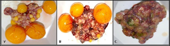
Fig.8.Normal and cancerous chicken ovary. (A) Gross anatomy of normal ovary with a hierarchy of developing follicles; (B) ovary classified as suspected; (C) ovary taken from a hen with metastatic late stage ovarian cancer;(Source: Erfan Eilati)
8.Avian Encephalomyelitis (AE)
AE is caused by vertically egg transmitted avian encephalomyelitis virus (AEV) and is a member of the Picornaviridae family . In young chickens, the neurological nature of the disease involves clinical signs, such as ataxia, paralysis, and tremors. Often clinical AE has been encountered in layer pullets following the application of live AE vaccine.
In laying birds, AE can negatively influence egg production and hatchability. Avian encephalomyelitis was diagnosed in breeding hens that experienced a transient decline in egg production (upto 20%). Additionally, a significant late embryonic mortality in the fertile eggs produced by these hens was reported. In a commercial laying flock, a dramatic fall in egg production (up to 75%), lasting for 2 weeks, was associated with encephalomyelitis. However, some naturally occurring outbreaks in non-vaccinated laying flocks showed lower rates (7.5% to 18%) of drop in egg production. The infected adult birds do not exhibit neurological disorders that usually appeared in the chicks hatching from eggs produced around and during the time of the egg drop. Virus replication in the ovary could be the most probable cause of the temporary decrease in egg production.
9.Fowlpox Fowl pox(FP)
FP is a common poultry viral disease and is characterized by cutaneous nodular lesions and/or fibro-necrotic lesions on the mucous membranes of the upper respiratory and/or digestive tracts. Avian pox viruses m(APV) are members of the Avipoxvirus genus in the family Poxviridae.
The decrease in the production of eggs in laying birds is as a result of the poor conditions of infected birds and the intensity of the proliferative lesions. Egg production may drop up to 15% in some flocks affected with FP.
10.Other Viral Infections
Avian hepatitis E virus (HEV) belongs to Orthohepevirus B species within the Hepeviridae family. In layer and broiler-breeder chickens, the virus has been associated with big liver and spleen (BLS) disease and hepatitis-splenomegaly (HS) syndrome. The two diseases have often been correlated with increased mortality (1–4%) and reduced egg production (10–45%) in broiler breeder and laying hens. Apart from the characteristic hepatosplenomegaly, oviduct regression is associated with production loss. In some breeder flocks infected with HEV, the hatchability was 36% to 46% lower than the expected level.
External parasites
There are a great number of external parasites that affect poultry by generating anemia, allergies, irritation, and secondary infections. Among these external parasites are lice (Menopon gallinae), fleas (Echidnophaga gallinacea), mites (Dermanyssus gallinae), flies, and ticks (Argas spp.). These parasites cause a decrease egg production as debility and loss of feed metabolism.
Helminths (internal parasites)
Helminths comprise internal parasites of three types: cestodes, nematodes, and trematodes. These worms invade different systems and organs and cause a wide range of lesions in laying hens. There is a large group of helminths that are parasites of the digestive system which affect the feeding processes of the poultry. As a result, laying is reduced. Among the most common examples of these parasites are:
- Nematodes: Ascaridia galli, Heterakis gallinarum, Capillaria.
- Cestodes: Raillietina
Coccidiasis
Coccidiasis is a subclinical disease characterised by mere presence of the coccidian organisms in the intestinal mucosa caused by protozoa of the genus Eimeria spp. and it will not cause clinical disease as coccidiosis. But, a marked effect on the digestive system of hens, preventing the correct absorption of nutrients; and cause drop in production.
Conclusions
In recent decades, intensive poultry production has faced both positive and negative consequences. Although there are several nutritional factors causing production problems, the bacterial and viral infections largely affect the reproductive organs or create severe health problems and production loss and their quality. The pathogens with a tropism for the reproductive organs or those that cause debilitating health conditions demonstrate mild to severe adverse effects on the egg production process. Multiple factors, mainly including host species, strain of the organisms, and age and immune status of the host, all play a major role in the type of reproductive disease. The systemic alterations caused by these organisms may have a negative influence on egg production and quality.
Using advanced techniques such as molecular analysis, microscopy, isolation and detection, would help in further understanding the pathogenesis of reproductive diseases caused by these infections. Further, an analytical and biological methodologies would help in understanding these diseases. Although there are a number of effective disease prevention protocols in place on poultry farms, still the bacteria and viruses continue to be a major constraint for the sustainability of egg production.
(Acknowledgements: The author thanks the google contribution of photos in this article)