The genus Eimeria is a protozoan parasite which causes acute invasion and destruction of intestinal mucosa. The Eimeria have many developmental stages in both inside and outside of the host. The oocysts usually oval, ellipsoidal, spherical/sub spherical in shape. There is a thining at the narrower pole called micropyle is present and these are covered with a cap like structure of different shape known as polar/micropyle cap. The sporulation of oocyst is depend upon the exposure to sun light, moisture, atmospheric temperature, types of soil etc. The time of sporulation of different species are varies from few hours to few days depends on the atmospheric temperature. The sporulated oocysts are identified on the basis of shape, size, presence/absence of micropyle and its cap, colour, form index, presence or absence of residual, polar and stiede bodies and time of sporulation. The coccidian oocyst, attaches to the epithelial lining in the intestines. It is a serious disease of animals develops in intestinal tract. The Eimeria species is much host specific parasite. The different Eimeria species such as; Eimeira apsheronica, E. arloingi, E. alijevi, E. caprina , E. hirci, E. christensini, E. jolchijevi, E. gilruthi, E. kochari and E. ninakohlyakimovae. Among the Eimeria species E. ninakohlyakimovae are most pathogenic species. Most commonly seen in kids below six month of age. Infection is more predominant in wet months. The parasite is transmitted oral route and infection may occur from residual contamination of the environment or from parasites being shed by adult goats.
Life cycle
The young one or kids ingest the oocysts, and parasite invades the gut epithelium. It divides asexually into a daughter cells in10-14 weeks, the daughter cells can multiply over a million fold. The daughter cells eventually break out of the gut and invade to a new area and repeating the process. In this stage the parasites attached to part of gut wall and developed into male and female gamonts. The females are fertilized and secrete oocysts into the gut wall and oocysts are then shed in faeces which completes the life cycle.
The young one pick up the infected oocysts in large quantity, produced by asymptomatic adults. The newly born kids pick up the infection during licking soiled dams, contaminated water, infected pasture.The infection may persist throughout the year but suitable time for disease spreading is usually onset of the monsoon where, proper temperature and moisture are available for sporulation of oocysts. Generally clinical disease seen post monsoon and winter season.
Symptoms
- Foul smelling faeces with or without blood streak
- Loss of appetite and severe constipation
- Loss of weight
- Severe abdominal pain
- Inappetance and anaemia
- Emaciation and unthriftiness
- Fever (ill kids lie down due to weakness and abdominal pain)
- Diarrhoea (stool in most severe condition includes bits of intestinal epithelium)
- Dehydration may aggravate the condition
- Sometimes death (in severe condition or untreated)
- The species are generally found in the small intestine, caecum and colon.
- Large numbers of oocysts may develop in the small intestine which may be thick white patches in appearance.
Diagnosis
- Identification of oocysts in faeces by salt/sugar flotation methods.
- Multiple species may be found in single samples.
- The number of oocysts found in the sample may depend upon the intensity of infection.
- Sometimes diagnosis may difficult from the symptom particularly in acute cases, where no shedding of oocysts in the faeces.
- For correct diagnosis, detection of typical gross lesions during post mortem examination is essential.
- The oedematous, thickened, haemorrhagic or congested excessive mucus is seen in entire intestine in acute cases.
- Pin point white cysts are found throughout the small intestine either deep or superficially on the closer examination of intestinal lining.
Treatment
- Treatment should be done with suitable anticoccidial drugs such as amprolium, sulphadimidine, monensin , sulfaquinoxaline, toltrazuril, lasalocid and decoquinate.
- Timely medication inhibits or checks the life cycle of the oocysts, and shorten the infection length, alleviate symptoms and may lessen the likelihood of reinfections as well as death.
- As life cycle of the coccidian is self-limiting and their life ends spontaneously, unless reinfection occurs in host.
- Infected animals should be removed and treated separately to prevent infection of others.
- Make sure of the treated animals are being treated effectively.
Prevention and control
- Provide good feeding practices including ample quantity of clean water.
- The sporulated oocysts may survive months to a year.
- Provide good managemental practices (i.e. sanitation).
- Provide proper space, avoid overcrowding.
- Sick animals should be separated from healthy one.
- The susceptible animals should be kept in clean and dry areas.
- Make sure neonatal receive colostrums properly.
- Provide proper treatment and care of sick animals.
- It is a common threat in kids
- Try to minimize the stress level.
- Equipments of feeding and watering should be kept clean, clear and free from any fecal contamination.
- Provide the ration containing coccidiostats (monensin, dequoqinate and lasalocid) which slow down the shedding of coccidian oocyst into the environment.
- The killing of oocysts can be done by heat, drying or direct sunlight.
- Bedding is the common source of infection.
- Imperfect diagnosis, resulting in heavy losses due to coccidiosis.
- The kid should not be allowed to graze/stay with adult goats.
- Screening and monitoring of animals should be done at regular interval.
- Provide ample quantity clean water.
- Avoid the mixing of kids and adult.
- Regular screening of coccidiosis should be done.
- Correct dose and timing should be preferred.
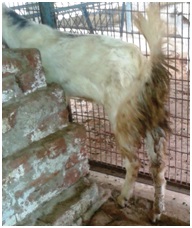
Goat showing Diarrhoea (typical indicator of coccidiosis) 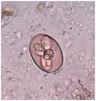
Sporulated oocyst 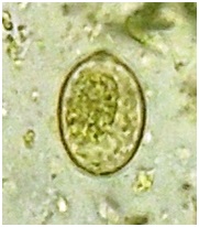
Unsporulated oocyst
Alok Kumar Singh1, Pradeep Kumar2, Amit Singh3 and J. Jayalakshmi4
1 Department of Veterinary Parasitology, College of Veterinary Science & A.H.,Kuthuliya, Rewa
2 Department of Veterinary Parasitology, DUVASU, Mathura
3 Department of Veterinary Parasitology, COVSc & A.H.,Kumarganj, Ayodhya
4 Department of Veterinary Parasitology NTR, COVSc., Gannavaram