Pallavi Khajuria, Gurpreet Singh2 and Dr. Amit Challana3
1 Assistant Professor, 2 Associate Professor, 3 Assistant Professor
Department of Veterinary Physiology and Biochemistry
Department of Veterinary Anatomy
College of Veterinary Science, Rampura Phul
Guru Angad Dev Veterinary and Animal Sciences University
India, as a tropical country, has a wide range of climatic zones, which make it vulnerable to a diverse range of parasites of veterinary and medical importance. Parasitic diseases pose a major health hazard in man and animal around the globe especially in tropical countries like India. Heavy economic losses are caused by ectoparasites to livestock industry due to their usual blood sucking habit that adversely affects the economic production.
Blood, being the most important specialized tissue of the body, creates an open channel system to provide the equal amount of nutrients, hormones and other important factors to different organs. Serum biochemical tests can be used for diagnosing various life threatening animal diseases which often result in severe economic losses in animals like reduced fur, wool and milk production. Variations in the serum biochemistry of infected animals depend on the species of worms. Biochemical parameters are very sensitive indicators for the degree of hepatic damage and the severity of parasitic infection.
Helminths are parasitic worms belonging to the lower invertebrate phyla of nematodes (roundworms) and platyhelminths (flatworms). Helminths are extremely diverse. They can be
classified into 3 taxonomic groups, that is, nematodes, trematodes, and cestodes, often with very different parasitic cycles.They may be transmitted orally (Strongyles,Fasciola sp., etc.), by transcutaneous route (Ankylostoma sp., Strongyloıdes sp., Schistosoma sp., etc.) by an arthropod vector (Onchocerca volvulus, Dirofilaria immitis, etc.).The definitive and intermediate hosts range from mammals,birds, reptiles to fish, molluscs and arthropods. They may be localized in organ lumen or in tissues such as gut, liver (parenchyma and bile ducts), lung, lymphatic vessels. Successive developmental stagesof parasitic species may infect different tissues and cells of different organs.
A wide variety of helminth species are able to colonise an extraordinary array of niches and host organisms, in each case circumventing host defence and expulsion mechanisms. The immunomodulatory abilities of helminths enables the worm to persist in the host and can lead to interactions with inflammatory and immune mechanisms involved in other infections or to vaccines or in allergic and autoimmune diseases. Interestingly, the strategy of helminths is not to outpace the immune system through rapid multiplication or antigenic variation, but to manipulate and modulate immunity in order to defuse immune defences, meaning the host fails to eliminate the parasites.
The most frequent gastrointestinal nematodes in cattle in are: Haemonchus sp. and Ostertagia sp., parasites of the abomasum Cooperia spp., Trichostrongylus spp., and Strongyloides spp., parasites of the small intestine Oesophagostomum spp., parasite of the large intestine.Gastrointestinal parasites are wide spread and appear in animals of all ages.
Changes in blood biochemistry in parasitic diseases
Infections can present a variety of clinical signs that range from no clinical manifestation at all to inappetance, diarrhea, vomition, anaemia and in severe cases, to death. Gastrointestinal nematodes produce also superoxide dismutase and glutathione S-transferase which neutralize toxic oxide radicals. A cystatin produced by Haemonchuscontortus and Nippostronylus brasiliensis modulates the antigen presentation to T cells by inhibiting cysteine proteases of antigen presenting cells, involved in the processing of the antigen. Biochemical effects are associated with reduced nutrient availability to the host through reduction in voluntary feed intake.
IgE levels are usually elevated in parasitized animals and many helminth infections (e.g. Oesophagostomosis, Ancylostomosis, Strongyloidosis, Bilharzia and Taeniosis) are associated with the characteristic type I hypersensitivity, such as eosinophilia, oedema and urticarial dermatitis. One of the best examples of IgE production and the allergies that result from it is the ‘self-cure’ phenomenon, which is seen in sheep infected with especially Haemonchuscontortus. Those worms that are embedded in the abomasal mucosa secrete antigens during their their moult. As a result, a local type I hypersensitivity develops locally; the combination of helminth antigen and mast cell bound IgE leads to mast cell degranulation and the release of vasoactive amines. These compounds stimulate smooth muscle contraction and increase vascular permeability, resulting in violent contraction of intestinal muscles together with an influx of fluid into the intestinal lumen. This combination results in the dislodgement of the worms and the major portion of the infection is expelled.
Inappetance with resultant reduction in dietary protein and plasma losses from damaged intestinal mucosa might be the main cause for hypoproteinemia. Inflammation of intestine by development stages of parasites might also because of poor absorption of protein metabolites resulting low level of total protein. Lowserum glucose in the diarrhoeic animals might be due to decreased appetite of animals, decreased absorption into the blood stream and rapid absorption and utilization of soluble carbohydrate and lipids from the gut by parasites.
Many of the helminths that infect their hosts percutaneously elicit a passive cutaneous anaphylaxis (PCA), a direct response to the antigens they liberate.Increased loss of endogenous protein into GI tract due to leakage of plasma proteins. Effect on gastrointestinal motility results in diarrhea, there by altering acid base balance. Reduction in nitrogen retention due to increased urinary nitrogen loss occurs implying reduction in the efficiency of utilization of absorbed aminoacids (e.g. Trichostrongylus colubriformis). pH mediated changes are also exhibited in abomasum in Ostertagia infection. There are changes in calcium and phosphorus absorption leading to reduced bone growth. In tapeworm infections like Monezia and metacestodes of Taenia spp intestines are virtually a solid mass of parasites resulting in obstruction and thus the animal exhibits symptoms such as unthriftiness, anorexia and diarrhea.
Cholecystokinin(CCK) is considered one of the most important factor in alimentary behavior. Parasites cause cellular damage in the intestine resulting in great release of CCK, thus reducing apetite.The migration of Cysticercus tenuicollis, cause traumatic hepatitis and death in young animals. Migration of cysticerci in the liver results in hemorrhagic and fibrotic tracts and serofibrinous peritonitis. White blood cells increase in number due to liver failure, inflammation following larval migration and stimulation of immune responses.Larval migration results in bleeding and congestions in the peritoneum and liver parenchyma due to reduction of RBCs, haemoglobin and hematocrit concentration. Biochemical parameters including liver enzymes (AST, ALT, ALP) and total bilirubin are increased and decreased total protein.
Fasciola gigantica produces superoxide dismutase which neutralizes superoxide radicals toxic for juveniles.F. hepatica releases cathepsin L-protease which cleaves IgE and IgG involved in the antibody dependent cellular cytotoxicity.
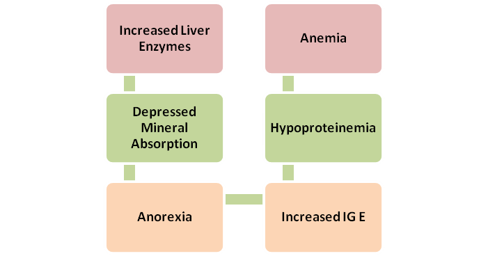
Thus the biochemical alterations caused by helminthic parasitism on host are usually associated with anorexia, loss of plasma proteins, alterations in protein metabolism, depressed levels of minerals, anaemia, pH mediated changes, depressed activity of intestinal enzymes and diarrhea.