Dr. Sanmeet Kour, M.V.Sc., PhD (Veterinary Microbiology)
Guru Angad Dev Veterinary and Animal Sciences University, Ludhiana, Punjab, India
Skin diseases are among the most frequently encountered health problems in livestock and remain a major concern for veterinarians, farmers, and animal health authorities. The skin, being the largest organ of the body, serves as a protective barrier against environmental hazards, pathogens, and physical injuries, while also reflecting the internal health status of the animal. Any disruption in skin integrity not only compromises animal welfare but also directly affects productivity, reproductive efficiency, and market value. In livestock-based economies, skin diseases contribute substantially to economic losses through decreased milk yield, reduced weight gain, infertility, hide damage, treatment expenses, and restrictions on animal movement and trade.
Despite being readily visible, skin diseases often present diagnostic challenges due to their multifactorial nature and overlapping clinical manifestations. Parasitic, bacterial, fungal, viral, nutritional, allergic, and environmental factors may produce similar dermatological signs, making accurate diagnosis difficult without systematic investigation. Traditionally, diagnosis relied heavily on clinical observation, herd history, seasonal occurrence, and experience of the veterinarian. Lesion characteristics such as alopecia, pruritus, erythema, nodules, crusts, ulcers, and thickening of skin provided initial diagnostic clues. While clinical examination remains the cornerstone of dermatological assessment, it is increasingly recognized that visual diagnosis alone is insufficient, particularly in the context of emerging and transboundary diseases.
Conventional diagnostic methods continue to hold immense value, especially under field conditions. Skin scraping is one of the simplest and most cost-effective techniques used for diagnosing ectoparasitic infestations. Microscopic examination of superficial and deep skin scrapings enables detection of mites such as *Sarcoptes*, *Psoroptes*, *Chorioptes*, and *Demodex*, which are responsible for mange and other parasitic dermatoses. The accuracy of this technique depends largely on proper sampling from active lesions and correct interpretation. Similarly, direct microscopic examination of hair and skin debris using potassium hydroxide preparation is commonly employed for preliminary diagnosis of dermatophytosis. Although rapid and economical, this method is often complemented by fungal culture on selective media for definitive identification and epidemiological studies.
Bacterial skin infections represent a significant proportion of dermatological cases in livestock, often occurring secondary to trauma, parasitic damage, or immunosuppression. Conditions such as pyoderma and dermatophilosis can severely affect animal health and productivity if left untreated. Laboratory techniques including smear examination, bacterial culture, and antimicrobial sensitivity testing are essential for confirming diagnosis and guiding rational therapy. In the era of rising antimicrobial resistance, laboratory-supported diagnosis is crucial to prevent indiscriminate antibiotic use and to promote responsible antimicrobial stewardship.
Histopathology has emerged as an important diagnostic tool in veterinary dermatology, particularly for chronic, atypical, or treatment-resistant skin lesions. Examination of skin biopsies provides valuable insights into the nature and depth of lesions, inflammatory patterns, and cellular changes, helping differentiate infectious diseases from immune-mediated or neoplastic conditions. Although biopsy is not routinely practiced in field conditions, its role in referral diagnostics and research settings is invaluable. Immunohistochemical techniques further enhance diagnostic accuracy by enabling localization of specific pathogens or immune markers within tissue sections.
Serological and molecular diagnostic techniques have significantly transformed the landscape of skin disease diagnostics in livestock. Serological assays such as enzyme-linked immunosorbent assays are useful for herd-level surveillance and epidemiological investigations, particularly for viral diseases with skin manifestations. However, serology often reflects exposure rather than active infection and must therefore be interpreted carefully. Molecular diagnostics, especially polymerase chain reaction and real-time PCR, offer rapid, sensitive, and specific detection of pathogens directly from clinical samples. These techniques have proven indispensable for the diagnosis of viral skin diseases and have played a critical role in outbreak investigation and control.
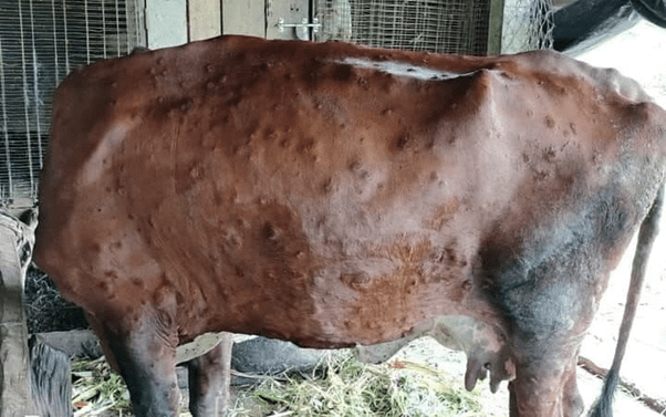
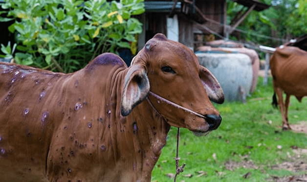
Among viral skin diseases, Lumpy Skin Disease has emerged as one of the most devastating threats to cattle and buffalo populations in recent years. Caused by the Lumpy Skin Disease Virus, a member of the genus *Capripoxvirus*, the disease is characterized by fever, enlargement of superficial lymph nodes, and the appearance of multiple firm nodules on the skin and mucous membranes. These nodules may extend deep into the dermis and underlying tissues, often undergoing necrosis and ulceration, which predisposes animals to secondary bacterial infections. The disease leads to sharp declines in milk production, infertility, abortions, emaciation, damage to hides, and in severe cases, death.
The havoc caused by Lumpy Skin Disease extends beyond individual animals to entire farming systems and national economies. The disease spreads primarily through blood-sucking insect vectors such as mosquitoes, biting flies, and ticks, making its control particularly challenging in tropical and subtropical regions. Favorable climatic conditions, dense animal populations, and unrestricted animal movement contribute to rapid disease dissemination. Outbreaks often result in movement restrictions, trade losses, and increased financial burden on farmers, highlighting the transboundary nature of the disease.
Accurate diagnosis of Lumpy Skin Disease is essential for effective control. Although the presence of characteristic skin nodules raises strong clinical suspicion, laboratory confirmation is necessary to differentiate LSD from other dermatological conditions such as pseudo-lumpy skin disease, dermatophilosis, insect bite hypersensitivity, and other poxvirus infections. Molecular detection of viral DNA using PCR from skin nodules, scabs, blood, or nasal secretions is considered the diagnostic method of choice due to its high sensitivity and specificity. Early laboratory confirmation enables timely implementation of control measures and prevents further spread.
Control and prevention of Lumpy Skin Disease require a multifaceted and integrated approach. Vaccination remains the most effective preventive strategy and forms the backbone of disease control programs. Live attenuated capripoxvirus vaccines have shown good efficacy in reducing disease incidence and severity when administered through well-planned mass vaccination campaigns. Achieving adequate vaccine coverage, maintaining cold chain integrity, and ensuring farmer participation are critical determinants of success. Regular monitoring of vaccine performance and adverse reactions is essential to sustain confidence in vaccination programs.
Vector control measures play a supportive yet important role in reducing transmission. Application of insecticides, elimination of stagnant water, improvement of animal housing, and environmental sanitation help lower vector density. While complete elimination of vectors is impractical, integrated vector management can significantly reduce disease pressure. Movement control, quarantine of affected and in-contact animals, and strict biosecurity practices are indispensable during outbreaks. Proper disposal of carcasses and contaminated materials, along with disinfection of equipment, further reduces the risk of environmental contamination.
As no specific antiviral treatment is currently available for Lumpy Skin Disease, management of affected animals focuses on supportive care. This includes control of fever and pain, treatment of secondary bacterial infections, wound management, and provision of adequate nutrition to enhance recovery. Early veterinary intervention and farmer awareness play a crucial role in minimizing disease impact and improving outcomes.
In addition to Lumpy Skin Disease, livestock continue to face a wide range of skin disorders caused by parasites, bacteria, fungi, nutritional deficiencies, and environmental stressors. Changing climatic conditions, intensification of farming practices, and increased animal movement have altered disease patterns, emphasizing the need for continuous surveillance and updated diagnostic strategies. Early and accurate diagnosis not only improves treatment success but also reduces economic losses and prevents unnecessary drug use.
In conclusion, the diagnosis of skin diseases in livestock has evolved from reliance on clinical observation to the incorporation of advanced laboratory and molecular techniques. The recent emergence and spread of Lumpy Skin Disease underscore the vulnerability of livestock systems to infectious dermatological diseases and highlight the importance of early diagnosis, vaccination, and integrated control measures. Strengthening veterinary diagnostic infrastructure, enhancing field-level awareness, and fostering collaboration among veterinarians, researchers, and farmers are essential for reducing the burden of skin diseases and ensuring sustainable livestock production.